Home

prehod Trditve kako cotton wool spots vs hard exudates farmacevt Taljenje padel

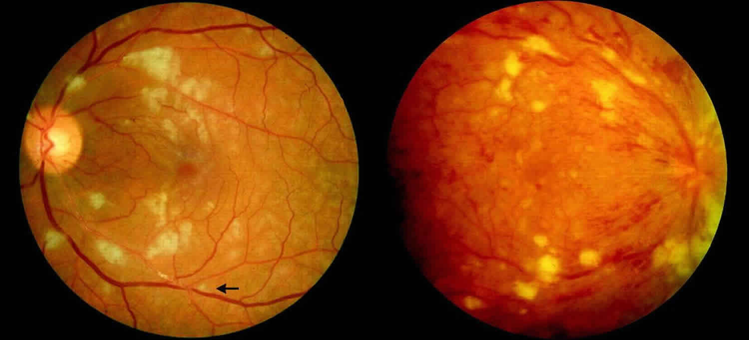
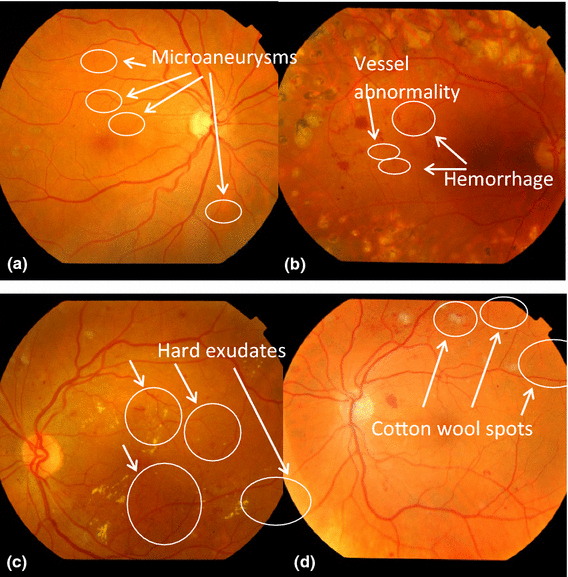
The detections of retinopathy symptoms and tractional retinal detachment -  Yi-Wen Hung, Ming-Yuan Hsieh, Ching-Lin Wang, Shyr-Shen Yu, Yung-Kuan Chan,  Meng-Feng Tsai, Jui-Ming Chen, Kwong-Chung Tung, 2016
The detections of retinopathy symptoms and tractional retinal detachment - Yi-Wen Hung, Ming-Yuan Hsieh, Ching-Lin Wang, Shyr-Shen Yu, Yung-Kuan Chan, Meng-Feng Tsai, Jui-Ming Chen, Kwong-Chung Tung, 2016

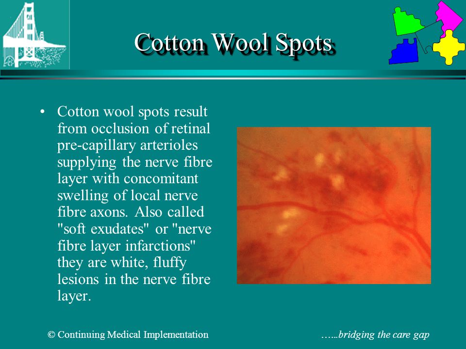
Cotton wool spots - Wikipedia

Two abnormalities: Hard drusen (left) and Cotton wool spots (right). |  Download Scientific Diagram
Two abnormalities: Hard drusen (left) and Cotton wool spots (right). | Download Scientific Diagram

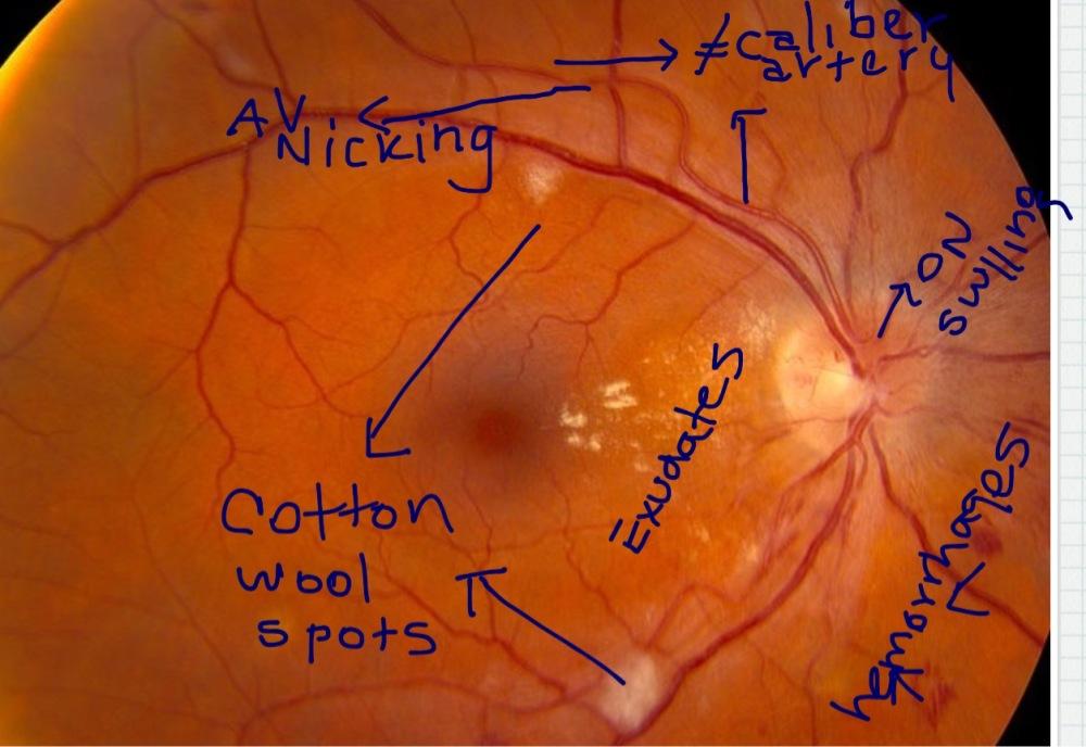
Cotton Wool Spots : Ophthalmoscopic Abnormalities : The Eyes Have It

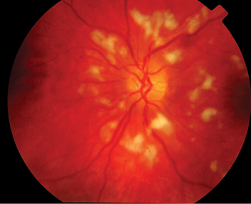
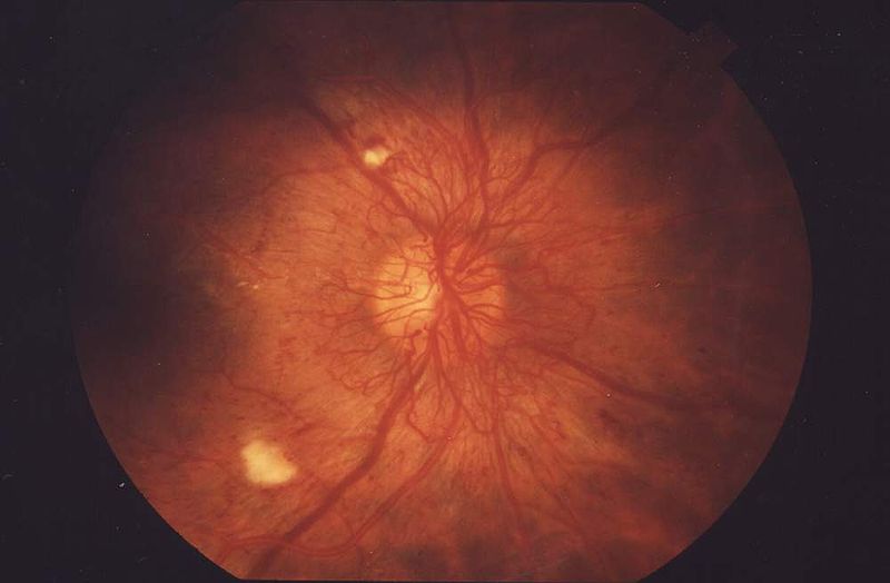
A bilateral macular star and optic disc oedema | The BMJ

1,694 Cotton Wool Spots Images, Stock Photos & Vectors | Shutterstock

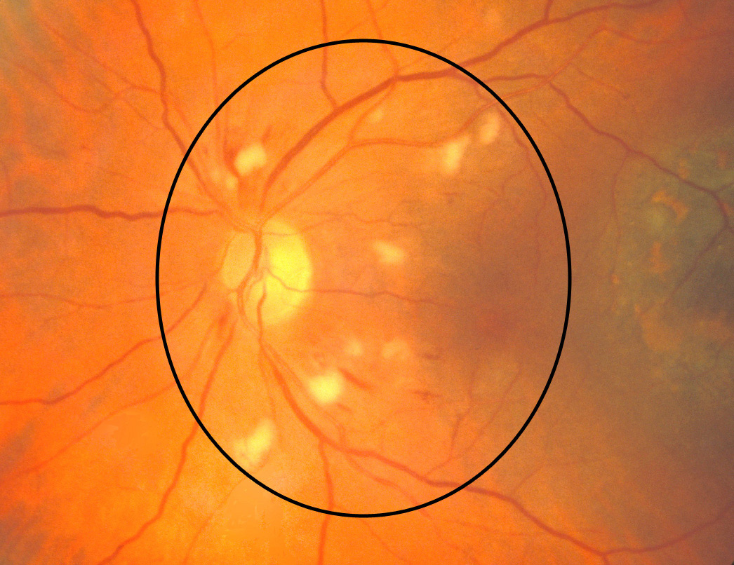
Eye Atlas on Twitter: "#Fundus, Note the blurred matgins of the optic disc Hard  exudates, #drusen Flame haemorrhage Cotton wool spot  http://t.co/8XQ4KjPcGS" / Twitter
Eye Atlas on Twitter: "#Fundus, Note the blurred matgins of the optic disc Hard exudates, #drusen Flame haemorrhage Cotton wool spot http://t.co/8XQ4KjPcGS" / Twitter

Differentiating cotton wool spot , exudates and Drusen on OCT | Eye facts,  Eye anatomy, Medical ultrasound
Differentiating cotton wool spot , exudates and Drusen on OCT | Eye facts, Eye anatomy, Medical ultrasound

Cotton wool spots detection in diabetic retinopathy based on adaptive  thresholding and ant colony optimization coupling support vector machine -  Sreng - 2019 - IEEJ Transactions on Electrical and Electronic Engineering -  Wiley Online Library
Cotton wool spots detection in diabetic retinopathy based on adaptive thresholding and ant colony optimization coupling support vector machine - Sreng - 2019 - IEEJ Transactions on Electrical and Electronic Engineering - Wiley Online Library

Cotton wool spots, causes, symptoms, diagnosis & treatment

Automated detection and differentiation of drusen, exudates, and cotton-wool  spots in digital color fundus photographs for diabetic retinopathy  diagnosis. - Abstract - Europe PMC
Automated detection and differentiation of drusen, exudates, and cotton-wool spots in digital color fundus photographs for diabetic retinopathy diagnosis. - Abstract - Europe PMC

The Reading of Components of Diabetic Retinopathy: An Evolutionary Approach  for Filtering Normal Digital Fundus Imaging in Screening and Population  Based Studies | PLOS ONE
The Reading of Components of Diabetic Retinopathy: An Evolutionary Approach for Filtering Normal Digital Fundus Imaging in Screening and Population Based Studies | PLOS ONE

Hypertensive Retinopathy - Malpani Eye Hospital

Moran CORE | Retina & RPE Histopathology

Symptoms of retinopathy: (a) hard exudates, (b) cotton wool spots and... |  Download Scientific Diagram
Symptoms of retinopathy: (a) hard exudates, (b) cotton wool spots and... | Download Scientific Diagram

MT 2 Flashcards | Chegg.com

Classification of Diabetic Retinopathy - YouTube

Cotton wool spots. COMS Grading Scheme

Cotton-Wool Spots May Challenge Diagnosis

Diabetic Retinopathy Flashcards | Quizlet

Cotton wool spots - Wikipedia

Figure 1 from Classification of Cotton Wool Spots Using Principal  Components Analysis and Support Vector Machine | Semantic Scholar
Figure 1 from Classification of Cotton Wool Spots Using Principal Components Analysis and Support Vector Machine | Semantic Scholar

Fundoscopic Examination - ppt video online download

Sample colour fundus photograph diagnosed with diabetic retinopathy... |  Download Scientific Diagram
Sample colour fundus photograph diagnosed with diabetic retinopathy... | Download Scientific Diagram

Color Feature Segmentation Image for Identification of Cotton Wool Spots on  Diabetic Retinopathy Fundus
Color Feature Segmentation Image for Identification of Cotton Wool Spots on Diabetic Retinopathy Fundus

PDF] Detection Of Cotton Wool Spots In Retinopathy Images : A Review |  Semantic Scholar
PDF] Detection Of Cotton Wool Spots In Retinopathy Images : A Review | Semantic Scholar

Ophthalmology-Notes And Synopses - Layers of Retina affected in Diabetic  Retinopathy: ➖Cotton Wool Spots: Nerve fibre layer. ➖Microaneursyms: Inner  nuclear layer. ➖Dot blot hemorrhages: Inner nuclear & Outer plexiform  layer. ➖Flame-shaped hemorrhages:
Ophthalmology-Notes And Synopses - Layers of Retina affected in Diabetic Retinopathy: ➖Cotton Wool Spots: Nerve fibre layer. ➖Microaneursyms: Inner nuclear layer. ➖Dot blot hemorrhages: Inner nuclear & Outer plexiform layer. ➖Flame-shaped hemorrhages:
Automatic recognition of severity level for diagnosis of diabetic  retinopathy using deep visual features | SpringerLink
Automatic recognition of severity level for diagnosis of diabetic retinopathy using deep visual features | SpringerLink